Easy Horse Hoof Anatomy
The hoof is perhaps the most fascinating structure of a horse. This relatively tiny foot supports your horse’s hundreds and hundreds of pounds on such a small frame. And then they propel your horse across the earth, sometimes with gusto. Horse hoof anatomy is mesmerizing and has many parts, each with a unique function.

This article does have two photos of the inside of the hoof as prepared specimens. They are at the VERY BOTTOM but you can jump to them here.
Jump to shopping for hoof supplies
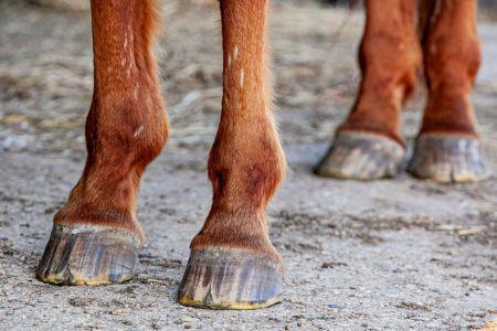
The outer hoof
The hoof wall
- What most of us think of as the hoof is what we can see – the hoof wall. And this outermost section has three distinct layers.
Stratum externum
- Horn cells make up this outer layer of the hoof wall. It’s a protective barrier between the world and the inner layers of the hoof.
The stratum medium
- Horn tubules running parallel to the hoof’s surface comprise the stratum medium, giving the hoof strength and durability.
Stratum internum
- Here’s where the magic happens. Live cells in the stratum internum make the horn tubules, keeping the hoof wall growing and regenerating. The new horn tubules form at the coronary band and proceed down the hoof. Slowly. Very slowly.
- The hoof wall, along with its friends the sole, frog, and heel bulbs, make up the hoof capsule.

The toe
- The front of the hoof is the toe. The toe’s length and balance significantly impact how a horse moves and can interfere with soundness.
- As a horse walks, it’s best for the heel to land first. Toe-first landings indicate hoof and lower leg issues and can cause long-term damage. Many horses with long toes run into problems, too, as this often goes hand-in-hand with underrun heels. Farriers can trim and round the edge of the toe to impact movement, too.
The quarters
- Quarters are on the sides of the hoof between the bulbs and the toe. The quarters are in the narrowest part of the hoof. The inner quarter closest to your horse’s midline is the medial quarter, while the outside quarters are lateral.
- The quarters should also be symmetrical, with the medial and lateral quarters being of equal size and shape.
- Sometimes quarter cracks occur, starting at the coronary band and running toward the hoof. These cracks can range from mild to openly bleeding, causing pain and lameness. Quarter cracks need adaptive shoeing, and sometimes your vet can surgically repair them.
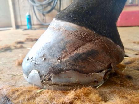
This is a healed quarter crack. This horse also has a pour-in shoe packing.
The coronary band
- Above the hoof wall lies the coronary band, also known as the coronet. The coronary band is the area of specialized tissue where the hoof wall meets the lower leg.
- The coronary band produces the hoof wall, as well as supplies nutrients and growth factors for other tissues of the hoof. A network of blood vessels and nerves in the area provides oxygen and nutrients to keep the hoof healthy and growing.
- Specialized epithelial cells produce the horn material that forms the hoof wall. These epithelial cells are fast to divide and multiply. Incidentally, the sole, frog, and bars of the hoof benefit from the coronary band as well. These fast-multiplying cells are the basis for the growth of these structures on the hoof’s underside.
- Daily inspections of the coronary band should reveal smooth tissue. You may find wounds, skin troubles like mud fever, or swelling. In laminitis cases, you may also see the hair above the coronary band starting to poke upwards. As laminitis progresses, the sinking of the bones pulls the hair in a new direction. This is not always the case with laminitis, but you may see it.
- When the coronary band is damaged from injury (even a small one) or disease, the hoof wall can grow abnormally or with defects. You may see rings around the hoof following a bout of sickness, laminitis, poor nutrition, or injury.
For more on the coronary band, read this gem.
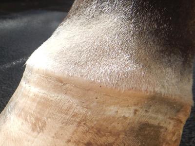
You can see the coronary band better on a clipped lower leg.
The Periople
- Below the coronary band lies the periople. This thin layer of tissue covers and protects the new horn growth from the coronary band. There is a visible texture to this layer, too. The periople does not cover the entire hoof wall, just the top portion.
- When the hoof is wet, the periople becomes softer and sometimes easier to differentiate from the hoof wall.
- Many farriers will rasp the entire hoof wall for that “show ring ready” look of the hoof. While this certainly makes the hoof look polished and tidy, there’s no reason to rasp the periople away, and some horses benefit from keeping the periople.
- Rats and mice are also known to eat the periople from hooves. Just one more reason to keep rodents away from the barn.

The periople is not always obvious. This horse hoof has a different texture, indicating the periople. Sometimes the farrier will rasp off this layer.
Inside the hoof
- Here’s where things get absolutely amazing! Who knew a few bones, squishy things, and cushions could hold up such a huge creature?
Bones of the hoof
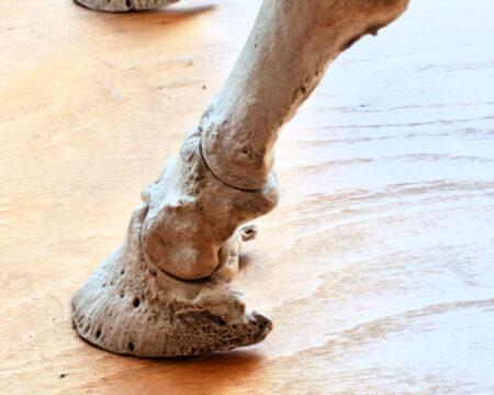
The coffin bone
- The coffin bone has many other names, including the pedal bone, the third phalanx, the distal phalanx, and P3. It’s the last bone in the horse’s leg, just below the short pastern bone. It also cups the navicular bone, which sits nestled at the back end of the coffin bone and the short pastern, towards the heels.
- Laminae connect the bone to the hoof wall. The porous bone surface gives soft tissue anchors for attaching laminae, tendons, and the digital cushion.
- The coffin bone helps to anchor some tendons, protects blood vessels, protects nerves, and assists with the break over of the hoof.
- When looking at the coffin bone, the bottom edge supports the horse’s weight. The wing of the coffin bone is along the back, where the deep digital flexor tendon attaches.
- As a horse ages, the coffin bone can change shape and density, and may fracture.
Amazing photos of the coffin bone can be found here.
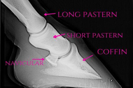
The navicular bone
- The navicular bone, also known as the distal sesamoid bone, is a small, flattened bone tucked behind the coffin bone. It rests between the short pastern bone and the deep digital flexor tendon.
- This small bone helps stabilize the hoof and helps your horse move smoothly.
- The navicular bone is notorious for being associated with swelling, degeneration, and breaks. Navicular disease, commonly referred to as “navicular” describes damage to this bone and its surrounding structures. Navicular disease can be painful and may benefit from corrective shoeing and other supportive care. It’s often tricky to diagnose, as a horse often suffers from both front limbs affected, thus making a horse appear sound.
The short pastern bone
- The short pastern bone, or the second phalanx or P2 bone, sits above the coffin bone and is mainly outside the hoof. It’s cylindrical and, like the coffin bone, is the attachment for tendons and ligaments.
- This handy little bone plays a big role in shock absorption and transferring weight throughout the limb. Ringbone affects the short pastern bone, causing bony growths that may circle the joints around the bone. Horses can also develop arthritis in this area.
Here you can see the P1, P2, and P3 (top to bottom)
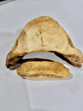
This is the coffin bone above the navicular bone, as if you were looking at the hoof from behind.

Other structures inside the hoof
Laminae
- The coffin bone is “attached” to the hoof wall with laminae inside the hoof. There are two types of laminae – the sensitive and non-sensitive laminae.
- The laminar layer is the meeting of finger-like projection from the coffin bone and the inner hoof wall’s surface. These fingers sort of “shake hands” to secure the bone to the hoof wall. There is also a bit of give to allow the hoof wall some movement.
- The inner surface of the hoof wall consists of the primary epidermal layer. This layer resembles many fingers and is layer upon layer of keratinized cells.
- The secondary dermal layer begins on the coffin bone’s outer surface. There are also finger-like projections here, although more of them and longer than the primary epidermal layer. Here the cells are more like skin cells.
- The laminae have a few critical functions in the horse’s hoof. Blood brings nutrients to the hoof, and the laminae provide structural support to the hoof and the leg. There is shock absorption by the laminae as a horse moves, too.
- During laminitis, this layer weakens, swells, and creates massive pain. The laminae may allow the coffin bone to rotate and drop if allowed to progress.
The digital cushion
- The digital cushion, sometimes called the plantar cushion, nestles below the coffin bone and between the frog and deep digital flexor tendon. The lateral and medial ungual cartilage flank the digital cushion, and the entire area is comprised of fiberous collage, fat, and connective tissue. The collagen gives structure, while the fat acts as a cushion.
- This squishy pad absorbs shocks and distributes a horse’s weight throughout the hoof. The overall shape of the hoof is supported, and the cushion helps with blood flow inside the hoof.
Ungual cartilage
- This supportive cartilage lies between the coffin bone and the hoof wall as a cushion as it extends towards the toe. The sole and frog are mostly comprised of this cartilage.
- This cartilage has a system of collagen fibers and elastin fibers, and is also rich in glycosaminoglycans. The collagen adds structure and strength, the elastin provides for stretching, and the glycosaminoglycans grease the joint surfaces.
The deep digital flexor tendon
- This hoof structure also exists outside of the hoof! The beginning of the deep digital flexor tendon (DDFT) is in the upper portion of your horse’s legs. The tendon travels down the leg, into the hoof, and attaches to the underside of the coffin bone. As the DDFT approaches the coffin bone, it gets wider.
- Blood vessels, nerves, and the synovial sheath, which holds lubricating fluid, cover the tendon.
- The DDFT serves to flex the horse’s lower leg. After a horse steps, the tendon pulls the horse’s leg backward, creating forward motion.
- In laminitis cases, this pulling force can out-muscle the damaged laminae and rotate the coffin bone.
The superficial digital flexor tendon
- Behind the DDFT is the superficial digital flexor tendon (SDFT). This tendon also originates high in the horse’s leg but attaches to the pastern bone and the short pastern bone inside the hoof.
- Like the DDFT, the SDFT flexes the lower leg and helps stabilize the hoof. It, too, is surrounded by nerves, vessels, and a synovial sheath.
The extensor digital tendon
- On the front side of the leg lies the extensor digital tendon (EDT). The EDT is the counterpart to the DDFT and the SDFT and extends the lower leg by lifting the hoof off the ground.
- Like the tendons at the back of the leg, it begins high on the horse’s leg and travels down into the hoof. The EDT attaches to the pastern, short pastern, and coffin bones. As with other tendons, it has the nerves, blood vessels, and synovial sheath, too.
Bottom of the hoof:
The sole
- This (sometimes) thick pad on the hoof’s underside is the sole. The sole cushions and absorbs shocks. The sole is comprised of keratinocyctes, which are the cells that make keratin. It’s this layer of keratin that forms part of the bottom barrier of the hoof.
- The sole has two parts – the peripheral sole and the central sulcus. As the name suggests, the peripheral sole is the outer portion. The central sulcus is the V-shaped groove down the center of the frog. The peripheral sole is thicker and tougher than the sulcus.
- The bars, white line, and frog are also part of the sole. And interestingly enough, the concave surface of the sole rarely connects with the ground.
- Horses with thinner soles often bruise easily and are sensitive to rugged and rocky surfaces. Protective shoeing and hoof pads can help.

This horse’s sole has a clear separation from the outer hoof wall.
The frog
- Within the sole and inside the V of the central sulcus is the frog. This squishy, almost rubbery, tissue aids in shock absorption and adds grip on slippery surfaces. The frog also distributes weight during movement and helps pump blood through the hoof.
- The frog should be level with the ground and the walls of the hoof, and thus should not require trimming unless there are flaking pieces.
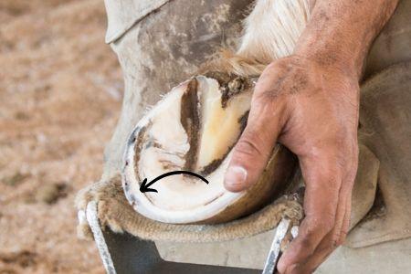
The collateral groove
- The collateral grooves are the channels running alongside the frog. These grooves help a horse move dirt and moisture out of the hoof. These grooves also help your farrier find the correct angle and length of the hoof wall.
- A horse with thrush often develops this bacterial infection in the collateral grooves. Movement, as well as topical medications, can help heal thrush.
The bars
- The bars are supportive structures running parallel to the hoof wall from the heel to the toe along the sides of the frog. Much like the hoof wall, the bars are dense and hard.
- The hoof capsule’s integrity is strengthened by the bars, and they also help with shock absorption. Much like other parts of the hoof, the bars benefit from regular trimming during a farrier visit.
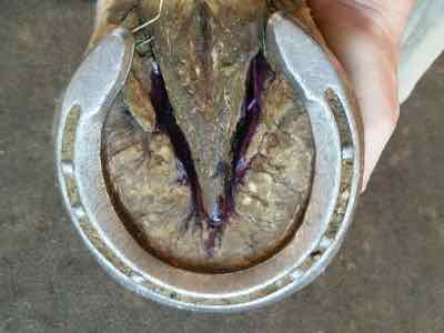
This horse’s frog is outlined by some purple thrush medication.
The heel bulbs
- The rounded, squishy structures at the back of the hoof are the bulbs of the heel, also called the hell bulbs. They rest on either side of the frog and extend back so you can see them without picking up the hoof.
- Not surprisingly, the bulbs provide shock absorption and cushion during movement. And like other softer parts of the hoof, the heel bulbs help circulation as they contract and expand during movement. Their texture and relative flexibility help on slippery surfaces, too.
The white line
- The white line, also known as the stratum medium of the hoof wall, is a visible line that appears on a horse’s hoof. Unless a horseshoe is covering it, and it’s not white. It’s more of a yellow color and delineates the junction of the outer hoof wall and the inner hoof parts. You can visualize the white line best on a freshly trimmed hoof where the hoof wall and the sole meet.
- It serves to protect the hoof from invasions by microbes. Unfortunately, cracks can create opportunities for the white line to become infected with bacteria. Hence, white line disease.
This second layer of hoof from the outside of the hoof wall is the white line. This horse’s white line is pretty close to white, and the fresh trim shows that.
The hoof as a second heart
- Horses don’t have muscles or soft tissue in their lower legs to send blood against gravity and back up to the heart.
- From the heart, blood pumps through the body via arteries. Blood returning to the heart travels through veins. The horse’s leg veins have one-way valves to prevent the blood from being pulled back down the leg. However, horses still need a way to push the blood against gravity through the veins.
- As horses move, their hooves act like a pump to push blood up the leg and toward the heart. This action is known as the hoof pump, hoof mechanism, or plantar pump.
- The hoof is seemingly rigid, but there is contraction and expansion as a horse moves. When the limb steps on the ground, the blood is pushed through the hoof as the blood vessels inside contract. As the weight is lifted, the hoof’s vessels expand, allowing gravity and the arteries to send blood into the hoof.
This barefoot horse has several large cracks.
14 Fun facts about horse hooves
- The keratin of the hoof is the same keratin that makes up hair.
- It’s a myth that white hooves are weaker than dark hooves. The only difference between the two is pigment.
- It can take several months for hoof supplements to impact hoof health.
- The horse’s closest living relatives also have an odd number of toes – the rhino and the tapir.
- Wounds to the coronary band can interfere with and permanently affect hoof growth.
- The hoof wall is 25% water.
- Some topical hoof treatments can harm hooves if the hoof wall is in poor condition. There is science about this here.
- The hoof can show damage, diet changes, and laminitic episodes as hoof rings. These markers will grow out as the hoof grows.
- The horse hoof takes about a year to grow out.
- Abscesses in the hoof sometimes pop out of the coronary band. This is known as gravel. You may find a wound around the coronary band. The abscess evacuation route may also turn into a horizontal crack.
- New evidence suggests that the other four toes of the horse’s precursors are absorbed by the hoof instead of becoming other structures, like splint bones, chestnuts, and ergots.
- The frog will sweat via specialized glands in the tissue.
- A horse’s metabolic health, specifically as it relates to insulin, is linked to hoof health. Higher insulin levels create damage in the hoof’s blood vessels.
- Thrush infections can happen to perfectly cared-for hooves in pristine environments as easily as horses in muddy conditions.

Stock up here for your horse supplies! As an Amazon Associate, I earn from qualifying purchases, but it’s ZERO extra cents to you. You can also visit my Amazon storefront here: PEG storefront.


If hooves are your jam, this book is for you.


and adds a nice sheen!


A simple and trusted hoof supplement.


For added squish during laminitis or other painful hoof trauma.


The gold standard for stinging hooves and anytime you need to pack the hooves.

Hi-quality no-bows with Back On Track reputation (that's good!)


These boots are the gold standard for jumpers and horses that like to interfere.


Support and softness for laminitis and other painful hoof conditions.


Buy bulk and save! This is great for stubborn hoof infections like seedy toe and thrush.

These squishy quilts are the perfect mix of no-bows and thicker quilts.


Feed your horse's hoof health from the inside out.

This supplement helps high-laminitis risk horses and has calming ingredients like magnesium.


The gold standard for bell boots in a rainbow of colors.


Classic, clean, stylish boots for your horse. Great for jumping.

Be practical when wrapping your horse's legs with a traditional color, but toss in some spice with the understated pattern!


Protect your horse's legs and let them shine bright.


These boots are my favorite for wrapping hooves with poultice or clay and a diaper. No more duct tape boots! You can also use inserts with these for more squish.


This style is great to protect the hoof, but is not soft and squishy for laminitis cases.


Show off your horse's legs with these safe, reflective, and fashionable boots.


These boots are great for protecting barefoot horses.


These SilverSox are great for protecting the legs from rubs, help with scratches and mud fever, and provide some compression, too.


Simple! Easy!


Take the heat out, reduce inflammation, help prevent long-term damage, and dress your horse in cute colors after a ride.

This is the strongest hoof pick available!

These are amazing for riding. They take some wrestling to get on, but they will stay on.
Thank you!
Here are some anatomy photos:

Can you spot the DDFT running under the navicular and connecting to the coffin bone?
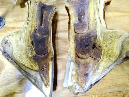
The hoof on the right has laminitis with rotation of the coffin bone. The hoof on the left is normal.